Colour me beautiful: photography at the nanoscale (Gallery thread)

Status
Not open for further replies.

Gaborn

Member
From a rusty microcanyon to a tie-dye leaf, these stunning images from the 2011 FEI Owner Image Contest use colour in inventive ways to transform snapshots from the microscopic world. New Scientist rounds-up the winners, runners-up and other striking entries.

0050651396d.jpg


Microcanyon

What appears as a deep crevasse is actually a mere microscopic crack in steel. Having subjected the metal to the rigours of a bend test, Martina Dienstleder of the Austrian Centre for Electron Microscopy and Nanoanalysis in Graz, decided that the results were pretty enough to photograph. Her colleague Manuel Paller then coloured the image with rusty tones and added a cloud-dotted sky to complete the look. The shot won the top prize.

(Image: Martina Dienstleder and Manuel Paller)
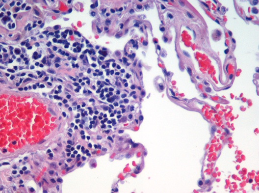
00506acbac2.jpg


Alveoli network

A dense network of blood vessels in the alveoli enables the quick transfer of oxygen to the bloodstream in the lungs. Here blood cells can be seen squeezing through the narrow capillaries. Taken by professional science photographer Oliver Meckes, the image won second prize in the competition.

(Image: Oliver Meckes)

00506dee4dd.jpg


Supersized spider

An arachnophobe's worst nightmare, a spider looms large in this image, taken using a scanning electron microscope. Also by Oliver Meckes. The colourised photo highlights the goliath's beady eyes and delicate hairs at 50-times magnification.

(Image: Oliver Meckes)

00506b456e0.jpg


Clam shell mite

Looking like a vibrant clam shell underwater, this vibrant image by Angelika Reichmann, also of the Austrian Centre for Electron Microscopy and Nanoanalysis in Graz, is actually a close-up of a mite's armoured body. Reichmann's colleague Margit Wallner coloured this photo, which claimed third prize.

(Image: Angelika Reichmann)
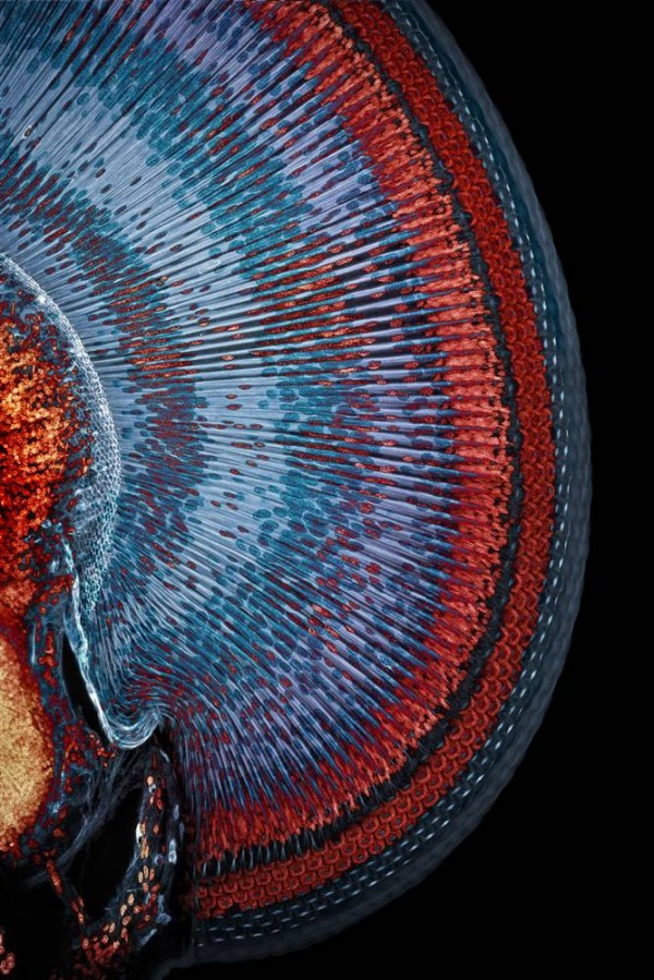
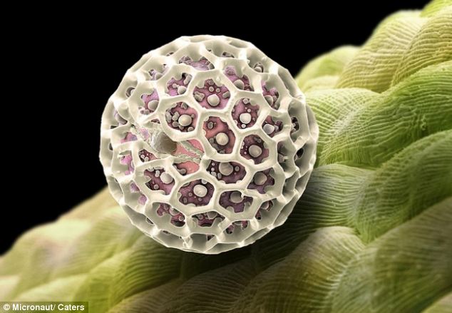
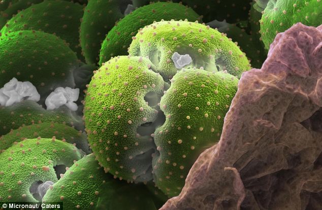
00506577a3e.jpg


Polyflower

This colourful shot by nanotechnology researcher Luca Boarino of the National Institute for Meteorological Research in Torino, Italy, resembles a close-up of a psychedelic sunflower. The delicate texture was created using nanospheres of polystyrene resin. Applied to a silicone surface, capillary forces caused the spheres to crack as they dried, completing the flower look.

(Image: Luca Boarino)
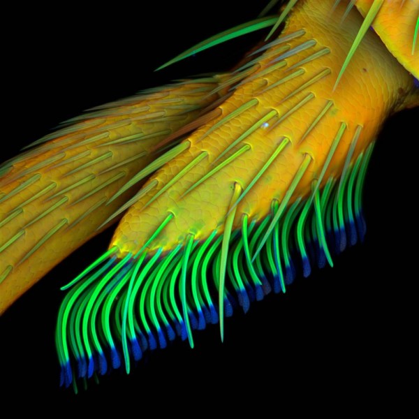
0050603363a.jpg


Tie-dye leaf

These tattered fibres are not from a ragged tie-dyed T-shirt, but rather show an abstracted cross-section of a leaf's veins. The rainbow image was created by Daisy Lee and Craig Queenan at the Nano-Structural Imaging Lab of Bergen County Technical Schools in New Jersey.

(Image: Daisy Lee and Craig Queenan)

005066650d9.jpg


Glacial shell

The icy blue tones of this micrograph transform the hard surface of a seashell into a chiselled glacier. It was taken by Wesller Schmidt, a technician at Universidade Federal de Minas Gerais in Belo Horizonte, Brazil.

(Image: Wesller Schmidt)

Gallery Here

I think it's neat how several of the images are COMPLETELY not what you'd think they were on first glance.
 
SneakyStephan said:
So if I understand it right most of these are manipulated by adding colors, right? (much like the hubble space images).

meh.
Well except for some of the shots (such as the insects ones) wavelengths of light that possess color cannot reach most of the objects depicted in microscopic photos. Most times they add color so that viewers can better distinguish what's in the photo.

Anyway more stuff!

Mouse skeletal muscle fibers
iom.jpg


Human sperm
img6.jpg


Velcro
img7.jpg
 
Metroid Killer said:
Well except for some of the shots (such as the insects ones) wavelengths of light that possess color cannot reach most of the objects depicted in microscopic photos. Most times they add color so that viewers can better distinguish what's in the photo.

How do they decide on the color? Is it an artist expression? Or is there some scientific method to decide which color it would be if the wavelenghts would reach it?

Awesome pics anyway.
 
Tence said:
How do they decide on the color? Is it an artist expression? Or is there some scientific method to decide which color it would be if the wavelenghts would reach it?
There aren't really any standards except that the coloring is very soft most often neon-like.

The reason to add color is really just to 'sell' the photos better. When you have to present some medical report concerning T-cells you better have some images which clearly shows the T-cell.
T-lymphocyte.jpg

Obviously T-cells aren't blue and you'll find them in all covers if you search on google. It's just to make them stand out and to present your ideas in a 'colorful' manner. They do try to make as much sense as possible though, or go with a theme. In the pollon shots above, notice how much green there are in the pictures solely because we are dealing with plants. And in the mouse muscle fiber shot, the actual fibers are colored red because we associate muscles with red.

Another way to capture color in microscopy is to use a fluorescence microscope. A sample is illuminated with light of a wavelength which causes fluorescence in the sample. The light emitted by fluorescence, which is at a different, longer, wavelength than the illumination, is then detected through a microscope objective. It still isn't the correct color as most of the samples are without color.

Fluorescence bloodcell
Bloodcell_sun_flares_pathology.jpeg


Yet another way to add color is to use stain or dye (limited to bulk tissue and cell populations) to enhance color contrasts in the microscopic images. You can say this method sometimes is more true in depicting the colors as it will enhance certain colors, other times the dye will just completely control what color the cells will have. Again it's up to each individual to decide which dyes to use, except for dyes that only work on certain sample. Some dyes have become standard for certain samples though (See Eosin for Red Blood Cells and muscle fibers).

Stained lung tissue
Emphysema_H_and_E.jpg
 
Macro fly
1246681253524204.jpg


Macro fly
1246681253524248.jpg


Macro leaf
b42b4c522766b5d77c4506cddfcfd839.jpg


Pollen
Pollen-from-a-Forget-me-not-petal.jpg


Soy Sauce
A-Microscopic-Snapshot-Of-Cancer-Cells-Multiplying-Photography_tc-n-_1.jpg


Salt and pepper
Everyday-Objects-Under-the-Microscope_Salt-Grains-and-Peppercorns1.jpg


Obviously the first 3 aren't true microscopic photos, but they're interesting none-the-less.
 
Seam said:
You mean in comparision with what you see in reality?
I think he's joking as pretty much all the images are colored in photoshop.

MOAR!

Caterpillar Mouth
IM_20110401_Waldron_74_caterpillar_lg.jpg


Sperm tails tangled up
IM_20100401_Barnes_2_Seminiferoustubule_lg.jpg


Mosquito eyes
IM_20100302_Meckes_3_Stechmuecke_450x_lg.jpg


Surface of eggshell
IM_20100707_Sharp_1_egg_shell_with_scale_bar_lg.jpg


Bidens Ferulifolia Pollen
IM_20100224_Hagen_1_Bidens_Ferulifolia_Pollen_lg.jpg
 
gorgeous images.

Hate to nitpick but these aren't really 'nano' scale, they're mostly micro (10^-6 meters). Things wouldn't be as outwardly interesting at the nanoscale with some context because the structures would be molecular/atomic.
 
Tiny friends!

Human flea
flea_1592351i.jpg


Tropical caterpillar
tropical-caterpill_1592360i.jpg


Dog flea
dog-flea_1592349i.jpg


Wasp
another-wasp_1592353i.jpg


Soldier turtle ant
soldier-turtle-ant_1592346i.jpg


Mites on mite
microscope-bugs-mite-covered-in-mites.jpg


Waterbear
waterbear.jpg


Pubic lice
25.jpg


6 days human embryo/egg
human-body-embryo.jpg


Most of these photos are by Steve Gschmeissner by the way. He has shot some really awesome microscopic images over the years.
 
xbhaskarx said:
What the hell is this thing...
Water bears can, among other things, survive temperatures of 303 degrees Fahrenheit and -460 degrees Fahrenheit, live for over 100 years without water, withstand over 1000 times more radiation than humans and survive ten days in outer space with no protection. Due to the small size of water bears, they have no need for any body systems that specialize in gas exchange. Therefore, they do not have a circulatory system or a respiratory system. When conditions get though, they basically die, stopping any process in their miniature bodies for up to 120 years! When conditions get better again, they revive and go on with their lives.

Cockroach bitches aint got nothing on these bears!

water_bear.jpg


C0089248-Water_bear,_SEM-SPL.jpg


C0069728-Water_bear_in_moss-SPL.jpg


C0069665-Water_bear-SPL.jpg
 
Metroid Killer said:
waterbear.jpg


Water bears can, among other things, survive temperatures of 303 degrees Fahrenheit and -460 degrees Fahrenheit, live for over 100 years without water, withstand over 1000 times more radiation than humans and survive ten days in outer space with no protection. Due to the small size of water bears, they have no need for any body systems that specialize in gas exchange. Therefore, they do not have a circulatory system or a respiratory system. When conditions get though, they basically die, stopping any process in their miniature bodies for up to 120 years! When conditions get better again, they revive and go on with their lives.

Wow.
 
Status
Not open for further replies.
Top Bottom